AI Dental Xray Analysis
Discover how artificial intelligence is transforming dental radiology — from diagnosis to documentation.
[
Learn How it Works
](#)
[
Compare AI Tools
](#)
[
Download AI Checklist
](#)
AI-Powered Radiographic Diagnostics
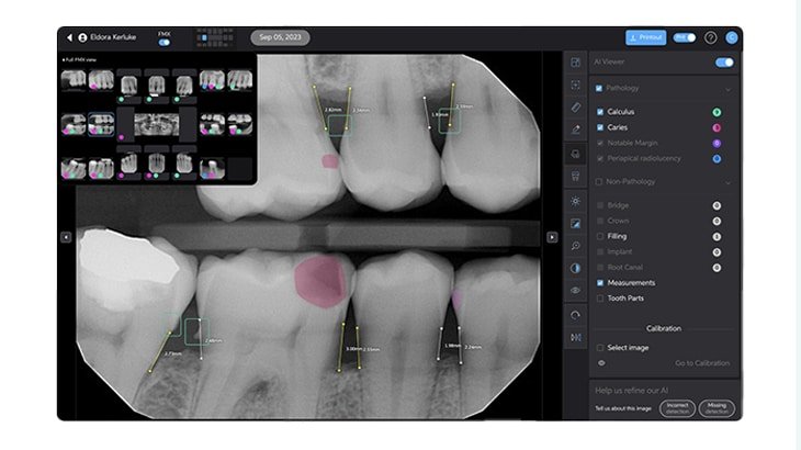
AI can detect up to 800 shades of gray in X-rays — the human eye sees only 30
Color-coded areas of concern allow for easy patient communication
FDA clearance proves an AI tool is safe, effective, and ready for clinical use — not just research or theory
Compare the Best AI Dental X-Ray Tools
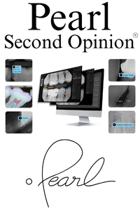
Pearl AI – Second Opinion®
Elevate Your Case Presentations
Pearl blends real-time diagnostic insights with visually intuitive overlays to help patients say “yes” to treatment. Show caries, calculus, and periapical issues with clarity — all in seconds.
Designed for dental teams focused on case acceptance and patient trust.
[
Read More About Pearl
](https://ai.dentist/ai-xray-detection/pearl-ai/)
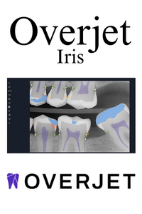
Overjet – IRIS Platform
Go Beyond Detection — Streamline Your Workflow
Overjet doesn’t just highlight caries and bone loss — it quantifies them, enhances image clarity, and ties it all to clinical performance and insurance workflows.
Built for practices that want diagnostic precision, patient clarity, and operational efficiency.
[
Read More About Overjet
](https://ai.dentist/ai-xray-detection/overjet-ai/)
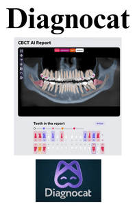
Diagnocat – AI Radiology Platform
All-in-One Diagnostic Intelligence
Diagnocat analyzes intraoral, panoramic and CBCT images and produces structured radiology reports and 3D models. Its US FDA clearances cover a CBCT second read for periapical radiolucency and 3D visualization; the reports themselves are not covered by those clearances.
Ideal for practices seeking CBCT analysis, per-tooth reporting, and seamless cloud-based collaboration.
[
Read More About Diagnocat
](https://ai.dentist/ai-xray-detection/diagnocat/)
Frequently Asked Questions
1. What is AI dental X-ray detection and how does it work?
AI dental X-ray detection uses advanced algorithms and deep learning to automatically analyze dental radiographs (2D and 3D images). It identifies abnormalities such as caries, bone loss, periapical lesions, and other pathologies by detecting subtle grayscale variations and anatomical patterns that may be missed by the human eye. The AI then highlights these findings visually on the images for dentists to review.
2. How accurate is AI compared to human dentists?
AI systems have demonstrated accuracy comparable to or exceeding that of experienced dentists in detecting dental conditions. They maintain consistent performance without fatigue or distraction, reducing human error and diagnostic variability. For example, AI can detect early caries and bone loss with high precision, improving diagnostic confidence.
3. Does AI slow down or speed up the diagnostic workflow?
AI significantly speeds up the diagnostic process by providing real-time analysis and automated annotations. This allows dentists to focus more on patient care and treatment planning rather than spending extensive time interpreting images manually.
4. Can AI detect conditions that are difficult for humans to see?
Yes. AI can detect subtle changes in grayscale and anatomical structures that may be imperceptible to the human eye, such as early-stage caries, minor bone density changes, or faint periapical radiolucencies. This early detection supports preventive and minimally invasive dentistry.
5. Is AI imaging FDA-cleared and clinically validated?
Many leading AI dental imaging tools, such as Overjet and Pearl’s Second Opinion, have received FDA clearance for specific diagnostic uses like caries detection and bone level measurement. This clearance ensures that the AI meets safety and efficacy standards for clinical use.
6. How does AI improve patient communication and case acceptance?
AI enhances patient education by providing clear, color-coded visual overlays on X-rays that highlight areas of concern. This objective evidence helps patients understand their dental conditions better, increasing trust and acceptance of recommended treatments.
7. Can AI integrate with existing dental imaging and practice management systems?
Yes. Most AI dental imaging platforms are designed to seamlessly integrate with popular imaging software and practice management systems, facilitating easy adoption without disrupting existing workflows.
8. Does AI replace the dentist’s expertise?
No. AI acts as a clinical co-pilot or second set of eyes, augmenting the dentist’s expertise by reducing oversight and providing consistent analysis. Final diagnosis and treatment decisions remain the responsibility of the clinician.
9. What types of dental images can AI analyze?
AI can analyze a variety of dental images including 2D bitewings, periapical, panoramic X-rays, and increasingly 3D CBCT scans. Advanced AI tools also provide quantitative measurements and enhanced visualization on these modalities.
10. How does AI handle continuous learning and improvement?
AI systems improve over time by learning from new data and cases, refining their algorithms to increase diagnostic accuracy and adapt to evolving clinical needs. This ongoing learning ensures that AI tools remain state-of-the-art.
These FAQs summarize the practical, clinical, and regulatory aspects of AI X-ray detection in dentistry, reflecting its growing role in improving diagnostic accuracy, workflow efficiency, and patient care.